

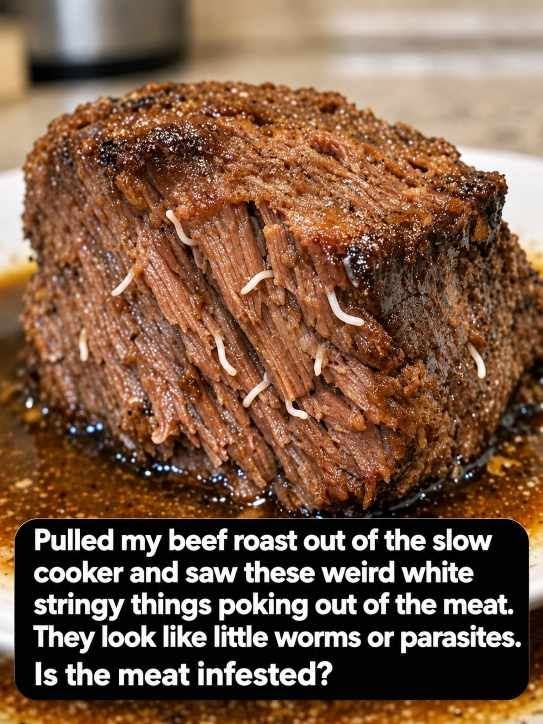
What Real Parasites in Beef Look Like
-
Cysticercus bovis (Beef Tapeworm Larvae): Cattle can occasionally act as intermediate hosts for Taenia saginata. However, tapeworm larvae do not manifest as long, smooth, branching tubes protruding from cooked meat. Instead, they form small, discrete, pea-sized fluid-filled cysts (often called "measly beef") embedded inside muscle tissue. These cysts are checked for rigorously by food inspectors at the slaughterhouse.
-
Trichinella spiralis: While historically feared in undercooked pork and wild game (such as bear or wild boar), Trichinella does not infect cattle, which are strict herbivores. Even in infected animals, Trichinella larvae are microscopic, measuring less than one millimeter in length and encysted within individual muscle cells, making them completely invisible to the naked human eye without a microscope.
-
Filarial and Roundworm Nematodes: While certain wild ocean fish frequently harbor visible roundworms (such as Anisakis), terrestrial cattle raised in regulated commercial agricultural systems do not host large, free-floating, macroscopic roundworms living inside deep, skeletal muscle tissue.
In modern commercial slaughterhouses, beef carcasses undergo strict veterinary inspections. Carcasses exhibiting signs of systemic parasitism or deep cysts are condemned and removed from the human food supply immediately. The odds of finding a macroscopic parasitic worm emerging from a commercially packaged supermarket chuck roast are vanishingly close to zero.
The Kitchen Detective: How to Verify What You Are Seeing
If you pull a roast from your cooker and need definitive proof that you are looking at vascular anatomy rather than an invasive organism, you can perform four simple sensory tests using a fork and your fingers:
-
Check for a Hollow Center (The Lumen): Slice across one of the white strands with a sharp paring knife. A blood vessel is an open biological pipe. Under good kitchen lighting or a magnifying glass, you will clearly see an open, circular lumen (the hollow interior through which blood once flowed). Parasitic worms, by contrast, possess solid internal organs and digestive tracts, lacking a clean, hollow, uniform structural pipe.
-
Observe the Branching Pattern: Trace the strand back into the roast with a fork. Blood vessels follow the natural dendritic laws of vascular systems: small capillaries merge into arterioles, which merge into larger branches. If you gently separate the meat grain, you will see the strand branching cleanly like the roots of a tree, firmly anchored into the connective framework of the muscle.
-
Test the Elasticity: Pinch the white strand between your fingers and pull gently. A blood vessel or elastin band will stretch with remarkable resistance and snap back into shape when released, feeling tough, smooth, and rubbery. A cooked worm or maggot, composed largely of delicate, coagulated insect or nematode protein, turns mushy, fragile, and disintegrates into a soft paste when pinched between two fingers after hours of boiling.
-
Note the Alignment with the Grain: Notice where the strands emerge. Blood vessels run parallel to the grain of the muscle fibers, as they are laid down alongside muscle bundles during the animal's physical development. They will never bore randomly across the grain or create irregular tunnels through the meat.
