

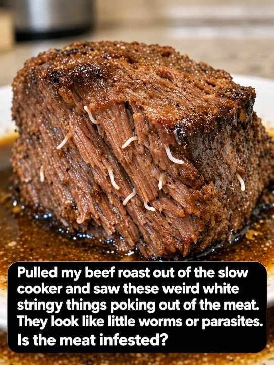
Few culinary moments cause as sudden a spike of panic as opening a slow cooker, lifting out a beautifully braised, tender beef roast, and spotting pale, curly, worm-like strands protruding directly from the muscle fibers. Against the deep mahogany brown of savory, shredded beef, these smooth, pale white tubes look unnervingly like parasitic nematodes, tapeworms, or maggots. For many home cooks, the immediate reaction is revulsion, followed by a rush to social media to ask whether an entire dinner is infested and must be thrown in the trash.
The reassuring truth is straightforward: that beef roast is not infested with worms, nor does it contain parasites.
What you are looking at is completely natural, sterile bovine anatomy—specifically, small blood vessels, nerve sheaths, or elastic connective tissue that have been pushed outward by the physics of slow cooking. While their pale color, tubular shape, and curled appearance mimic foreign organisms to the untrained eye, their presence is an expected byproduct of how tough cuts of meat behave when braised for hours.
Understanding why these white strands appear requires an exploration of bovine muscular architecture, the differing thermal properties of protein fibers, the true nature of meat parasites, and simple tests you can perform in your own kitchen to put your mind entirely at ease.
The Verdict: Anatomy, Not Infestation
When a piece of beef like a chuck roast, brisket, shank, or bottom round spends six to eight hours in a slow cooker or Dutch oven, the muscular landscape transforms dramatically. The mysterious white strings that pop up between the grain of the meat are arteries, veins, or elastin-rich fascial conduits.
Every living muscle in a steer is heavily vascularized. In life, muscle fibers require a constant, pressurized supply of oxygenated blood and nutrients, as well as a network of returning veins and nervous signaling pathways. These vessels travel in intricate branches throughout the entire muscle body, running parallel to and weaving between the individual muscle fiber bundles.
Under normal, raw conditions, these vessels are flat, translucent, or filled with microscopic fluids, blending seamlessly into the pink, marbled mass of raw beef. It is only after extended cooking, moisture loss, and muscle contraction that these structures become visible, white, and prominent, poking out in the exact manner that triggers alarm.
The Anatomy of a Pot Roast: Collagen vs. Elastin
To understand why these strands remain firm and pale while the surrounding beef turns brown and shreddable, one must examine the two primary structural proteins found in animal connective tissue: collagen and elastin.
Tough cuts traditionally chosen for pot roasts come from well-exercised, load-bearing parts of the animal, such as the shoulder (chuck), chest (brisket), or rear leg (round). These hard-working muscles are fortified by massive networks of connective tissue designed to withstand continuous tension and physical load.
The Behavior of Collagen
Collagen makes up the overwhelming majority of the connective tissue encasing muscle fibers (the endomysium, perimysium, and epimysium) and attaching muscle to bone via tendons.
Chemically, collagen is composed of a tight, triple-helix protein structure. When exposed to dry, rapid heat (such as a quick sear on a grill), collagen contracts aggressively, squeezing out moisture and turning meat leathery and tough. However, when collagen is subjected to gentle, low, moist heat over several hours—typically between 160 degrees Fahrenheit and 205 degrees Fahrenheit (71 to 96 degrees Celsius)—a transformative hydrolytic reaction takes place:
-
The water molecules in the cooking liquid penetrate the triple helices.
-
The hydrogen bonds holding the collagen chains together unravel and break apart.
-
The tough, fibrous collagen converts into gelatin, a luscious, melt-in-the-mouth liquid that enriches the braising liquid, coats the tongue, and gives pot roast its signature tender, succulent texture.
